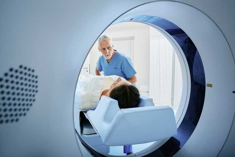
Why have I have been referred for a three-dimensional (3D) standing CT image?
Traditionally, imaging of the foot and ankle involves plain x-rays, which are two-dimensional (2D), and the standard images taken are from above and from the side. It’s then up to your clinician to estimate how these would look in 3D.
As technology has evolved, it is now possible to obtain a 3D scan in a standing functional position using Standing CT. It gives your doctor a much better picture of what might be wrong and helps them better diagnose and treat your health concern. A Standing CT scan is particularly helpful for diagnosing issues with flattened arches, bunions or other toe deformities.


